Product Details
Place of Origin: CHINA
Brand Name: AVACARE
Certification: ISO13485, CE0123
Model Number: 2.5mm
Payment & Shipping Terms
Minimum Order Quantity: 1000 units
Price: negotiable
Packaging Details: Individually packed by blister pouch, 10 units/inner box, 200 units / carton
Delivery Time: 20-30 working days
Payment Terms: L/C, T/T
Supply Ability: 300000units / month
Name: |
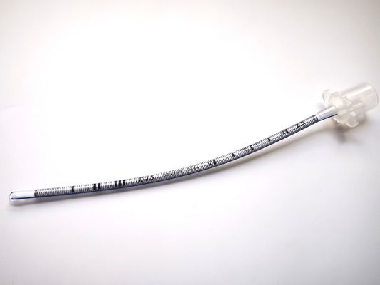
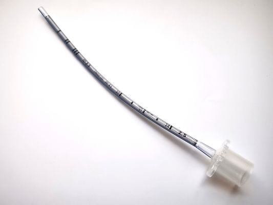
Reinforced Endotracheal Tube Uncuffed |
Material: |
PVC |
Parts: |
X Ray Line, Murphy Eyes |
Type: |
Medical Grade |
Size: |
2.5mm |
Use: |
Respiratory Medicine |
Name: |
Reinforced Endotracheal Tube Uncuffed |
Material: |
PVC |
Parts: |
X Ray Line, Murphy Eyes |
Type: |
Medical Grade |
Size: |
2.5mm |
Use: |
Respiratory Medicine |
Description:
HENAN AILE INDUSTRIAL CO., LTD is a company for operating medical disposables, and the Reinforced Endotracheal Tube is one of our productions.
The Reinforced Endotracheal Tube, which has Material reinforcing spiral within the wall of the entire tube, is suitable for both oral and nasal intubation. And it is a method of inserting a special endotracheal tube into the trachea or bronchus through the mouth or nasal cavity.
X-ray endotracheal intubation is a medical procedure that uses fluoroscopic imaging to guide the placement of an endotracheal tube into the trachea. It is performed to ensure accurate and safe placement of the tube, particularly in challenging cases or when visualization of the vocal cords is difficult.
Procedure:
Patient Preparation: The patient is positioned supine with the neck extended and the head in a neutral position. The oropharynx is anesthetized with a topical agent to reduce discomfort and facilitate intubation.
Equipment Setup: The fluoroscopy machine is positioned so that the patient's neck and chest are clearly visible on the monitor. The endotracheal tube is selected based on the patient's size and anatomy.
Tube Insertion: The laryngoscope is used to visualize the vocal cords, and the endotracheal tube is advanced through the vocal cords and into the trachea under direct vision.
Fluoroscopic Visualization: Once the tube is in place, fluoroscopic imaging is used to confirm its position. The tube should be positioned just below the vocal cords, with the tip in the mid-trachea.
Securing the Tube: The endotracheal tube is secured in place using tape or other appropriate devices to prevent accidental dislodgement.
Benefits:
Accurate Placement: X-ray guidance allows for precise placement of the endotracheal tube, reducing the risk of malposition or esophageal intubation.
Real-Time Visualization: Fluoroscopy provides real-time visualization of the airway and the position of the tube, enabling immediate correction of any malposition.
Reduced Complications: X-ray endotracheal intubation helps reduce the risk of complications such as esophageal intubation, pneumothorax, and airway trauma.
Useful in Difficult Cases: X-ray guidance is particularly beneficial in challenging cases, such as patients with difficult anatomy, obesity, or cervical spine abnormalities.
Indications:
X-ray endotracheal intubation is indicated in the following situations:
Anticipated difficult intubation
Patients with cervical spine instability or trauma
Patients with anatomical abnormalities of the airway
Patients with a history of difficult intubation
Obese patients
Patients with limited neck mobility
Contraindications:
X-ray endotracheal intubation may be contraindicated in certain situations, such as:
Patients with unstable vital signs
Patients with severe respiratory distress
Patients with a known allergy to contrast agents (if used)
Conclusion:
X-ray endotracheal intubation is a valuable technique that enhances the safety and accuracy of endotracheal tube placement. By using fluoroscopic guidance, healthcare professionals can visualize the airway in real-time and ensure that the tube is positioned correctly, reducing the risk of complications. It is particularly useful in challenging cases where direct visualization of the vocal cords is difficult or when there is a high risk of malposition.
Product composition and function of Reinforced Endotracheal Tube Uncuffed:
Total Size(mm) | 3.0/3.5/4.0/4.5/5.0/5.5/6.0/6.5/7.0/7.5/8.0/8.5/9.0/9.5 | |
15mm connector | Reliable connection to all standard equipment | |
Wire coil | Increasing flexibility, providing effective resistance to kinking | |
Murphy Eye | Reducing the risk of occlusinon and maintaining airflow |
2.5mm Application Features Reinforced Endotracheal Tube Uncuffed:
It is designed to reduce the risk of kinking when a patient’s head is in an extended or flexed position.
It is suitable for both oral and nasal intubation.
The Reinforced Endotracheal Tube Uncuffed also has Tip-to-Tip X-ray line, which allows for safe positioning control.
Material reinforcing spiral within the wall of the entire tube helps to prevent the tube from kinking.