Product Details
Place of Origin: CHINA
Brand Name: AVACARE
Certification: ISO13485, CE0123
Model Number: 3.5mm
Payment & Shipping Terms
Minimum Order Quantity: 100 units
Price: negotiable
Packaging Details: Paper-plastic bag packaging, sterilization, small packaging, 100 units/inner box, 200 units / carton
Delivery Time: 20-30 working days
Payment Terms: L/C, T/T
Supply Ability: 300000units / month
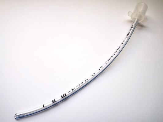
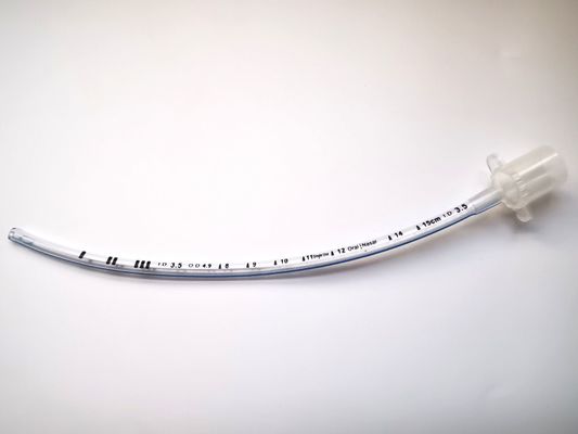
Name: |
Standard Endotracheal Tube Uncuffed |
Material: |
Pvc |
Parts: |
X Ray Line, Murphy Eyes |
Type: |
Medical Grade |
Size: |
3.5mm |
Use: |
Endotracheal Tube |
Instrument Classification: |
Class II |
Shelf Life: |
3 Years |
Cuff: |
No |
Name: |
Standard Endotracheal Tube Uncuffed |
Material: |
Pvc |
Parts: |
X Ray Line, Murphy Eyes |
Type: |
Medical Grade |
Size: |
3.5mm |
Use: |
Endotracheal Tube |
Instrument Classification: |
Class II |
Shelf Life: |
3 Years |
Cuff: |
No |
HENAN AILE INDUSTRIAL CO., LTD is a company for operating medical disposables ,our main products are specialized in anesthesia products and respiratory products . In detailed, the anesthesia products include Nasal Endotracheal Tube, Preformed Oral/Nasal Endotracheal Tube,Reinforced Endotracheal Tube.
Amoung them, the Nasal Endotracheal Tube is a method of inserting a special endotracheal tube into the trachea or bronchus through the mouth or nasal cavity. And the Nasal Endotracheal Tube Uncuffed is one type of it, which has different size to adapt to different medical needs,including 2.0mm to 10.0mm.
A preformed endotracheal tube is a type of endotracheal tube that comes with a specific curvature or shape designed to facilitate easier insertion and alignment with the anatomy of the airway. These tubes are pre-shaped during manufacturing to match the natural curvature of the trachea and vocal cords.
The use of a preformed endotracheal tube can offer several advantages, including:
Total size of Endotracheal Tube Uncuffed (mm) | 2.0/2.5/3.0/3.5/4.0/4.5/5.0/5.5/6.0/6.5/7.0/7.5/8.0/8.5/9.0/9.5/10.0 | |
Application Features | 1.Suitable for both oral and nasal intubation. | |
2.Tip-to-Tip X-ray line allows for safe positioning control. | ||
3.Murphy eye incorporated as an additional safety feature. |
During intubation, a physician usually stands at the head of the bed looking towards the patient's feet and with the patient lying flat. The positioning will vary depending on the setting and whether the procedure is being done with an adult or child. With children, a jaw thrust is often used.
The endotracheal tube with the assistance of a lighted laryngoscope (a Glidescope video laryngoscope is particularly helpful for people who are obese or if a patient is immobilized with a suspected injury to the cervical spine) is inserted through the mouth (or in some cases, the nose) after moving the tongue out of the way.
The scope is then carefully threaded down between the vocal cords and into the lower trachea. When it's thought that the endotracheal tube is in the proper location, the doctor will listen to the patient's lungs and upper abdomen to make sure that the endotracheal tube was not inadvertently inserted into the esophagus.
Other signs that suggest the tube is in the proper position may include seeing chest movement with ventilation and fogging in the tube. When a doctor is reasonably sure the tube is in position, a balloon cuff is inflated to keep the tube from moving out of place. (In infants, a balloon may not be needed). The tube is then taped to the patient's face.
Once the tube is in place, it's important to verify that it is truly in the proper location to ventilate the patient's lungs. Improper positioning is particularly common in children, especially children who have experienced trauma.
In the field, paramedics have a device that allows them to determine if the tube is in the correct position by a color change.In the hospital setting, a chest X-ray is often done to ensure good placement, though a 2016 review suggests that a chest X-ray alone is inadequate, as is pulse oximetry and physical examination.
In addition to directly visualizing the endotracheal tube pass between the vocal cords with a video laryngoscope, the authors of the study recommended an end-tidal carbon dioxide detector (capnography) in patient's that had good tissue perfusion, with continued monitoring to make sure the tube does not become displaced.
In the setting of a cardiac arrest, they recommended using ultrasound imaging or an esophageal detector device.